
Cellular responses to
tissue injury
Abstract—
1. Introduction01Edge overlap
aControl
bTreatment
cBaseline
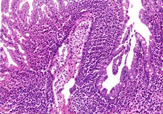
dRecovery
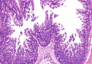
2. Results03
aControl
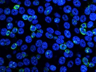
bVehicle
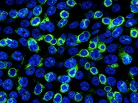
cTreatment
3. Discussion
Target
123456
37 kDa
Identify image duplication within manuscripts and across published literature, with traceable evidence and expert review.
Data-protection measures during transfer and processing.
Link source materials and review records for traceability.
Retention and deletion follow the agreed service terms.

















From image extraction to expert review, with traceable evidence at every stage.
Examine local duplication and image correspondence after cropping or geometric transformations.

Compare tissue structures and local textures to locate duplicated regions in histology images.

Examine cell distributions and local reuse across migration and invasion assay fields.

Compare band morphology and local backgrounds to locate duplicated bands or lanes.

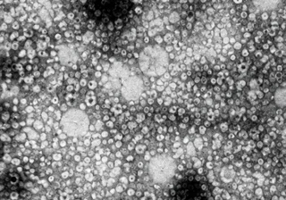
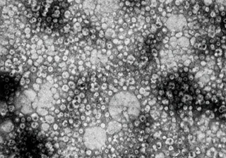
Compare ultrastructure and texture to assess correspondence after cropping or transformation.

Mask quadrant lines, axes and text to examine correspondence between scatter distributions.

Compare cell distributions and field structure to identify possible reuse across experimental groups.

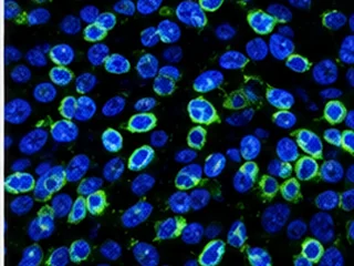
Examine single-channel and merged images to distinguish documented relationships from candidate duplication.

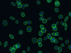
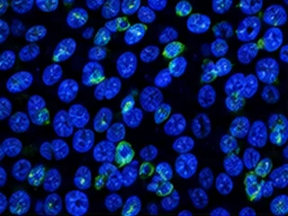
Compare imaging locations and spatial signal distributions to identify repeated fields and local reuse.
Biomedical and AI research underpin our image integrity screening.
 Shanghai University





 Tsinghua University
 Zhejiang University
 Peking University
Institution logos scroll automatically. Hover or focus to pause; leave to resume. Press Space to pause or resume.
Patents and software copyrights support continued research and product development.
Peking University Research Center
FigScan's professional image-screening system helps our laboratory prevent research misconduct and improve research quality. Its straightforward, intuitive operation has significantly improved our efficiency.
Nature Biotechnology
FigScan is a great addition to the peer-review process, greatly improving the efficiency and accuracy of plagiarism detection.
Qilu Hospital, Shandong University
As medical researchers, we use FigScan to help ensure the originality of the images in our papers. Its detailed and accurate findings make it a dependable research assistant.Zhejiang University School of Medicine
FigScan provides a highly professional service, particularly for medical image duplication screening. Its user-friendly interface and ease of use make it a valuable assistant in our medical research.Testimonials scroll automatically. Hover or focus to pause; leave to resume. Press Space to pause or resume.
Reviewers examine image comparisons, captions and experimental groups to distinguish documented insets and same-field channel relationships from potential duplication. Complex cases receive additional analysis within the agreed scope, with review notes retained.
Supported research imagery includes fluorescence, flow cytometry, Western blots, histology, H&E, electron microscopy, microscopy and wound-healing assays. Accepted file formats and submission limits are specified in the screening console.
Similarity scores indicate image correspondence; they cannot establish the nature of an issue by themselves. Findings must be reviewed alongside original images, experimental groups and captions. Reports provide technical evidence and review notes, not determinations of research misconduct.
The Image Integrity Review Report presents image locations, corresponding regions, similarity scores and review notes. Services that include literature searches also provide published-image matches. Deliverables depend on the selected service.
Institutional services can be tailored to screening volume, participating teams, confidentiality requirements and delivery format. Contact channels are listed at the bottom of this page.


